Hémorragies maculaires du myope
Les myopes saignent volontiers en rétro-fovéolaire.
C’est souvent associé à une membrane néovasculaire qui nécessite un traitement rapide, qui sera vite efficace : des injections d’anti-VEGF
Mais il n’y a pas toujours de membrane néovasculaire.
Parfois ils saignent spontanément.
Ces hémorragies spontanées vont régresser spontanément en quelques semaines.
Spon-ta-né-ment
Et sans cicatrices ni fibrose … Ni traitement
Ce sont des ruptures de la membrane de Bruchs avec rupture de la choriocapillaire
Un article met en évidence comment les distinguer.
‘Structural OCT changes distinguishing between myopic macular haemorrhages due to choroidal neovascularization and spontaneous Bruch’s membrane rupture: the « myopic 2 binary reflective sign »
Cecilia Mularoni et al.
Eye (Lond). 2023 Oct 9. doi: 10.1038/s41433-023-02780-w. Online ahead of print.
L’étude
C’est une étude portant sur 47 patients
· 13 avec une hémorragie spontanée
· 34 avec une membrane néovasculaire
Les auteurs ont trouvé deux signes OCT (d’où le terme de « binary ») permettant de les différencier :
- Une lésion hyper-réflective homogène au niveau de la rétine externe
- Une ligne hypo-réflective homogène entre l’hémorragie et l’épithélium pigmentaire
Cette ligne hypo-réflective est le signe de l’absence de membrane néovasculaire, donc qu’il ne faut pas injecter.
Ça ressemble à quoi ?
A ça :
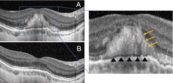
A : la lésion à l’origine
B : régression spontanée en 6 mois
Flèches jaunes : matériel hyper-réflectif
Triangles noirs : ligne hypo-réflective séparant l’hémorragie de l’EP
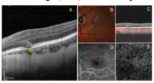
Flèches jaunes : on retrouve la ligne hypo-réflective : ce n’est pas une membrane, il n’y a pas d’injection à faire
C’est confirmé par l’OCT-A
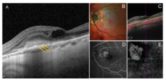
Flèches jaunes : PAS ligne hypo-réflective, mais une ligne hyper-réflective : c’est une membrane, il faut injecter rapidement
C’est aussi confirmé par l’OCT-A
C’est statistiquement significatif
· Sensibilité 100%
· Spécificité 97%
· Valeur prédictive positive 93%
· Valeur prédictive négative 100%
Donc une très belle fiabilité pour identifier les patients qui ont cette hémorragie spontanée, comme ceux qui sont à traiter
Abstract
Objective:
To evaluate the sensitivity and specificity of structural optical coherence tomography (OCT) in comparison to fluorescein angiography (FA) and OCT angiography (OCTA) in discerning between macular haemorrhages (MH) due to myopic choroidal neovascularization (m-CNV) and idiopathic macular haemorrhage (IMH) in myopic patients and to suggest a new OCT biomarker to discern these two entities.
Methods and analysis:
In this longitudinal retrospective study, patients affected by MH and pathological myopia were included. All patients underwent OCTA and FA to discern bleeding from m-CNV or IMH. Furthermore, all patients underwent a structural OCT and 2 expert graders evaluated the presence of the myopic 2 binary reflective sign as a biomarker to discern between IMH and bleeding from m-CNV.
Results:
Forty-seven eyes of 47 patients were enrolled. By means of angiographic examinations, 34 out of 47 eyes with MH (57%) were diagnosed as m-CNV, whereas 13 eyes (43%) as IMH. Using structural OCT, the graders identified the presence of the myopic 2 binary reflective sign in 13 out of 13 eyes with IMH. In 33 out of 34 cases with m-CNV, the 2 graders established the absence of the sign. This accounted for 100% of sensibility and 97% of specificity of structural OCT in discerning between MH from m-CNV and IMH.
Conclusion:
Structural OCT can discern with good reliability between IMH and bleeding from m-CNV based on the presence/ absence of the myopic 2 binary reflective sign. This could be of paramount relevance in the clinical setting for the diagnosis and treatment of HM patients.
|
|

