BMC Ophthalmol. 2023
Un nouvel acronyme : CHAFES
L’équipe d’Alan Bird a publié en juillet 23 un nouvel acronyme
Les anglosaxons doivent donner un prix pour les acronymes, tellement il y en a ! Voici donc un acronyme de plus : CHAFES.
· CRSC
· Hypermétropie
· Axiale (comme longueur axiale courte)
· F pour Folds : plis choroïdiens
· Elargie/épaissie : les couches oculaires
· Scléral pour Modifications sclérales
On connaissait déjà la coexistence des yeux courts et hypermétropes.
L’association avec la CRSC a déjà été décrite.
Les plis choroïdiens sont rares, mais assez fréquents en cas d’hypermétropie
Ils décrivent un signe nouveau (en tous cas pour moi) : un épaississement des « enveloppes oculaires » à l’échographie. (On ne fait pas assez d’échographie). Entendons la sclère.
Ils montrent aussi un aplatissement du pôle postérieur, au lieu d’être uniformément arrondi, décrit comme « le signe T ». On a déjà vu un pôle postérieur aplati dans des sclérites. Mais ce n’est pas le cas ici.
La combinaison de toutes ces choses ENSEMBLE n’a été décrite nulle part
Ce groupe anglais a trouvé 7 cas de patients présentant tous ces signes et hop !, un acronyme ! Ceci décrit, ils ne savent pas comment l’expliquer. Est-ce que la sclère est coupable ? La choroïde ? Ils ne savent pas.
On retient :
- Que la présence de plis chorio-rétiniens doit nous imposer une échographie oculaire
- Que l’important dans tout ça est la prise en charge de la CRSC, car ce sont des patients en âge de travailler qui sont à risque de passer doucement en cécité fonctionnelle.
Imagerie multimodale du cas 2 :
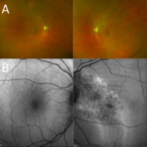 A- Image Optos montrant des plis choroïdiens dans la région maculaire, prédominant à gauche
B- Auto-fluorescence montrant un aspect de CRSC chronique, avec des altérations gravitationnelles de l’EP dans l’œil gauche, et des plis chorio-rétiniens
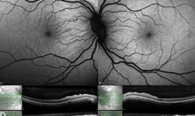
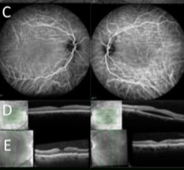 C- ICG montrant les plis chorio-rétiniens bilatéraux
D- OCT à l’inclusion montrant le fluide sous-rétinien à gauche apparaissant au cours du suivi
E- OCT vertical lors du suivi montrant des plis
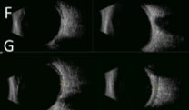 F- B-scan montrant les pôles postérieurs aplatis des deux côtés
G- B-scan montrant l’épaississement des couches oculaires de l’œil gauche
Pour en savoir plus
Combined central serous chorioretinopathy, hypermetropia, short axial length, chorioretinal folds, enlarged/thickened ocular coats, with varying association of scleral changes (CHAFES) Susan M Downes, Sonia P Mall, Saoud Al-Khuzaei et al. BMC Ophthalmol. 2023 Jul 14;23(1):322. doi: 10.1186/s12886-023-03038-5. Affiliations expand PMID: 37452273 PMCID: PMC10349429 DOI: 10.1186/s12886-023-03038-5
Abstract
Purpose: To describe a condition with the following features: chronic central serous chorioretinopathy (CCSC), chorioretinal folds, scleral changes (including any of the following flattened or ‘squared off’ posterior pole, ‘T sign’, or thickened ocular coats), accompanied by a short axial length and hypermetropia in a series of 7 patients.
Methods: The case notes of 7 patients presenting with a combination of CSC, choroidal folds scleral changes and hypermetropia were reviewed as part of a retrospective case series. Corrected visual acuities, serial refraction, colour imaging, fluorescein and indocyanine green angiography findings, together with B-ultrasound scan features were recorded, with axial length measurements as available (< 23.3 mm was defined as short).
Results: The study included 14 eyes of 7 subjects (2 females and 5 males) with a primary presentation of central vision disturbance. All patients showed signs of previous or current episodes of the following features in at least one eye: CSC (5/7 bilateral); choroidal folds (6/7 bilateral), thickening of ocular coats in the 5 in whom this was measured, at least one scleral abnormality on ultrasound in at least one eye. A short axial length at final appointment was recorded in 13/14 eyes.
Conclusions and relevance: The combination of CCSC with choroidal folds, hypermetropia with apparent shortening of the eyeball associated with one or more scleral abnormalities such as a flattened or ‘squared off ‘appearance of the B ultrasound may be a specific ocular condition. The aetiology of this particular combination of posterior segment manifestations is unknown; the choroid could be the primary focus of disease with secondary involvement of the sclera. Alternatively, the features observed may result from a chronic inflammatory process affecting the sclera with secondary effects on the choroid, retinal pigment epithelium and retina. In our case series, the final vision was not significantly different from vision at presentation.
