Découverte

Pachychoroïdes : une 7è entité

Une septième entité découverte en 2022 ; mélange entre des néovaisseaux de type 1 (occultes) et le SPP.

Le 17 mars 2024

Le spectre des Pachychoroïdes recouvre 6 entités :

1- La CRSC (choroïdo-rétinopathie séreuse centrale)
2- L’EPP (épithéliopathie pigmentaire et pachychoroïde)
3- Néovascularisation pachychoroïdienne (NVP)
4- Vasculopathie Polypoïdale Choroïdienne (VPC) ou dilatation anévrysmale de type 1
5- Excavation focale choroïdienne (EFC)
6- Syndrome de pachychoroïde péri-papillaire (SPP)

Non seulement toutes ces pathologies ont en commun la pachychoroïde, mais en plus les patients peuvent passer de l’une à l’autre !

Par exemple, une EPP est une forme chronique et/ou frustre de CRSC/

En 2022, une nouvelle entité a été découverte : un mélange entre des néovaisseaux de type 1 (occultes) et le SPP

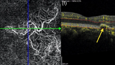
Le Syndrome de pachychoroïde péri-papillaire (SPP) est définie comme du liquide sous et intra-rétinien en nasal de la fovéa, dans l’aire péri-papillaire. Cette effusion est due à une hyperperméabilité choroïdienne au pourtour de la papille.

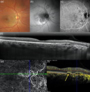
Dans ce cas, il y a un SPP + des néovaisseaux. Ça nous fait une nouvelle abréviation : « NPP » pour néovascularisation pachy-choroïdienne péri-papillaire.

En fait, plus on regarde les choroïdes, plus on trouve de syndromes….


Pour en savoir plus

Peripapillary pachychoroid neovasculopathy: A novel entity Javier Montero Hernández et al.

Eur J Ophthalmol. 2022 Jan;32(1):NP149-NP153. doi: 10.1177/1120672120953071  Epub 2020 Aug 25.

Abstract

Purpose:

Peripapillary pachychoroid syndrome is a new variant of pachychoroid disease recently described. It is important to establish the features and pathogenic mechanisms of this novel entity and its possible association with type 1 neovascularization. The aim of this study is to report a case of peripapillary pachychoroid neovasculopathy, a novel entity of pachychoroid spectrum disease.

Case report:

A 51-year-old woman was referred to our macula service due to macular pigmentary changes in her right eye. On examination, best corrected visual acuity was 20/20 Snellen equivalent in both eyes. Funduscopy revealed pigmentary changes in the papillonasal bundle in her right eye with mottled autofluorescence in the same area where pigmentary changes. Spectral-domain optical coherence tomography illustrated a shallow irregular pigment epithelium detachment (PED) which appears as “double layer sign” overlying pachyvessels, with hyper-flow signal beneath it and a large network of neovascularization revealed by cross-sectional optical coherence tomography angiography. Indocyanine green angiography confirmed the pachyvessels and choroidal hyper-permeability in the peripapillary region. A diagnosis of peripapillary pachychoroid neovasculopathy was made and good visual and anatomical outcomes were achieved with a treat-and-extend regimen with intravitreal aflibercept.

Conclusion:

We report a novel entity within the pachychoroid spectrum disease, the peripapillary pachychoroid neovasculopathy, which could improve our understanding of the pathogenic mechanism of choroidal neovascularization in pachychoroid disorders.